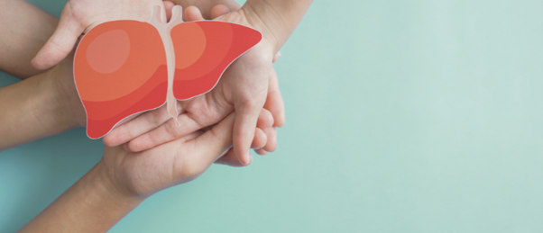
Developing 3D-bioprinted liver tissue models to study common liver disease
In a bid to gain further understanding of the etiology and pathogenesis of metabolic dysfunction–associated steatohepatitis (MASH), a team of researchers has created 3D bioprinted liver models.
MASH, previously referred to as nonalcoholic steatohepatitis, is an inflammatory condition characterized by the accumulation of fat in the liver, which can lead to cirrhosis and liver failure.
With no cure for the disease, organ transplantation is currently the only option for treatment. Ongoing research efforts exploring potential therapeutic approaches to resolve steatohepatitis have been hampered by the lack of preclinical models that fully encapsulate the intricacies of the condition.
Seeking to address this issue, researchers from Sanford Burnham Prebys, Viscient Biosciences, the Salk Institute and the University of California, San Diego (all CA, USA) created 3D bioprinted liver tissue models. These were produced by layering human hepatocytes and nonparenchymal liver cells, including Kupffer cells and liver sinusoidal endothelial cells.
The cells were sourced from donors with healthy livers and MASH-diseased livers. The resulting product was a MASH-diseased tissue model that exhibited fibrosis, a healthy liver tissue model and a chimeric tissue model composed of both healthy cells and diseased nonparenchymal cells.
 Infographic: Comparing preclinical models of non-alcoholic steatohepatitis
In this infographic, we compare four preclinical models: in vitro 2D models, in vitro 3D spheroid models, in vivo animal models and in vitro organ-on-a-chip models, highlighting the pros and cons of each, in the context of investigating the liver disease non-alcoholic steatohepatitis.
Chimeric models revealed the role of hepatic stellate and liver sinusoidal endothelial cells in the disease pathogenesis. By observing increased collagen deposition and increased secretion of extracellular matrix proteins, the team concluded that both cell types played a role in promoting fibrosis in the 3D bioprinted tissue model.
Discussing the team’s approach, co-author of the study and Chief Scientific Officer of Viscient Biosciences, Jeffery Miner stated “We believe this advance enhances the translation of our results to human clinical trials and drug discovery. This model represents a fully human system with the potential to detect clinically active targets and therapies. That’s important given that current discovery and animal models have not translated into any approved drugs.”
With MASH affecting an estimated 30% of the world’s population, this development marks a significant breakthrough for liver disease research. The study highlights the importance of developing 3D human liver tissue models that allow researchers to gain further insight into the complexities of MASH.