New open-source microscope shows brain–behavior connection in real time

Researchers have developed an open-source microscope that can track a freely swimming zebrafish while also imaging fluorescently labeled cells in its brain.

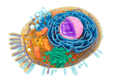
Credit: Barth van Rossum
Discerning the link between brain activity and behavior is a particularly tantalizing challenge for neuroscientists. Testing that connection in a living organism in real time is difficult, but imaging brain activity at the cellular level in a freely moving animal has been a nearly impossible task.
Now, researchers in Germany have developed a microscope system that allows them to do just that—in transgenic zebrafish expressing fluorescent calcium indicators. “This approach makes it possible to observe neuronal activity during unrestrained behavior. We can test the larvae in different environmental conditions and can immediately analyze the effects,” said senior author Gil Westmeyer, from the Helmholtz Zentrum München and the Technical University of Munich, in a press release.
Westmeyer’s team designed their NeuBtracker microscope using commercially available components. The system has two cameras: an infrared-sensitive camera for locating the fish in a water-filled “arena” and a fluorescence-sensitive camera that tracks the fish with the help of galvanometric mirrors and software written by the researchers. The entire arena can be illuminated by an LED light for fluorescence excitation, and a fast-refocusing, electrically tunable lens ensures good resolution for fluorescence imaging of the whole fish or just its head.
“It is finally possible to see the effects of pharmacological substances on the behavior and the neuronal activity—or other cellular signal processing events—at the same time and across an entire organism,” said Westmeyer of the system’s capabilities.
The researchers took advantage of their modular design to demonstrate two different configurations for the microscope: MicroFixed, which incorporates a fixed-magnification lens for the fluorescence camera, and MacroZoom, which incorporates a zoom lens for adjustable magnification. The latter design includes additional lasers that could be used to produce different illumination patterns across the arena, create visual cues for the fish, provide photostimulation, or potentially even control gene expression optogenetically. In addition, different arena designs can be fabricated by 3-D printing, including vessels with reservoirs for drug delivery or multi-well plates for high-throughput screening.
Using the NeuBtracker, Westmeyer and his colleagues demonstrated that fluorescence in the hindbrain and in cerebellar granule cells correlated with larval swimming speed. They went on to visualize neuronal activation and behavioral changes in free-swimming zebrafish larvae exposed to the odorant cadaverine, the neurostimulant 4-aminopyridine, or repeated light-dark illumination cycles.
“The selective expression of fluorescent sensor proteins allows us to detect the activity of particular neurons,” said Panagiotis Symvoulidis, the study’s first author.
The new system also has the advantage of being open-source, with a parts list, blueprints, and software all available on the NeuBtracker website. “We wanted to give our scientific colleagues the possibility to build their own NeuBtracker because we had been waiting for such a device for years,” said Westmeyer, who sees great promise in the new microscope. “This systemic approach enables us to make new discoveries and we will, for example, seek to use this device in drug discovery and metabolic research.”