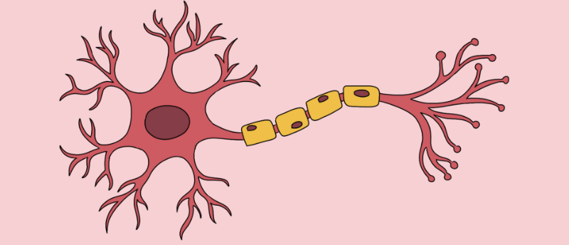
Visualizing nerves in vivo using photoacoustic imaging
Researchers demonstrate the suitability of photoacoustic imaging to non-invasively visualize and differentiate myelinated nerves from surrounding tissue.
Surgery and other invasive medical procedures that require anesthetic or nerve blockades can cause peripheral nerve injury, which leads to long-lasting sensory and motor problems. Researchers at Johns Hopkins University (MD, USA) have characterized the absorption and photoacoustic profiles of myelinated nerves across the near-infrared spectrum to show that multispectral photoacoustic imaging can be used as a non-invasive interoperative technique to prevent nerve injury during surgery.
During surgery, nerves can be mistaken for other tissues and accidentally cut, stretched or compressed. To mitigate the risk of nerve damage, ultrasound and MRI scans have been trialed to assist surgeons in locating nerves during a procedure. However, it is challenging to distinguish nerves from the surrounding tissue using ultrasound, and MRI is a costly and time-consuming technique.
Multispectral photoacoustic imaging is a promising non-invasive technique to capture detailed images of tissues in the body. First, the target region is illuminated with pulsed light, which causes the tissue to heat up and expand slightly. This emits ultrasonic waves that are picked up by an ultrasound detector.
 Get in formation: ultrasound waves direct cells into place
How do you precisely move something you can’t see with your own eyes, let alone touch?
In this study, the research team determined the ideal wavelengths for identifying peripheral myelinated nerve tissues with photoacoustic imaging. Myelin is a protective layer that forms around neurons and contains lipids. The researchers hypothesized that wavelengths between 1630–1850 nm would be optimal as the lipids in the myelin sheath have a characteristic absorption peak in this range.
They performed optical absorption measurements on peripheral nerve samples from pigs and observed an absorbance peak at 1210 nm, which is also present in other types of lipids. When the contribution of water was subtracted from the spectrum, a unique nerve tissue peak at 1725 nm was observed.
The researchers also conducted photoacoustic measurements in vivo in pigs, using a custom imaging setup. This confirmed that the lipid absorption peak at 1725 nm is characteristic of nerve tissues. The results also indicate that the 1630–1850 nm range can be used to differentiate lipid-rich nerve tissues from other types of tissues, materials containing water or those that are lipid-deficient.
Senior author Muyinatu Bell concluded, “our results highlight the clinical promise of multispectral photoacoustic imaging as an introspective technique for determining the presence of myelinated nerves or preventing nerve injury during medical interventions, with possible implications for other optics-based technologies.”