Discovering biomarkers in a dish
 Amina Ann Qutub is a biomedical engineer at the University of Texas at San Antonio (TX, USA) who studies the design of human cells and develops computational models to predict how single cells form neural networks and to screen novel biomedicines. Amina also launched the Quantu Project, which aims to understand how our daily behaviors affect the brain’s ability for cellular repair. Here, Amina discusses her research and how transformative networking has been throughout her career.
What is the main focus of your research?
My research focuses on developing computational tools and experiments to uncover how our cells communicate, what happens when this communication goes wrong and how we can correct cell communication to treat disorders and regenerate tissue. I am particularly interested in how cells of our brain and vascular system communicate.
What approaches do you use to model the brain for biomedicine?
Many! Live imaging, cell culture, cell engineering, molecular sequencing, human behavior tracking and computer modeling, among others. One focus of my research is understanding how neurons develop and change in response to our environment and genetics. For this, we designed an approach called ‘Living Neural Networks’, to study the growth and fate determination of neural cells over time, spatially and functionally over weeks.
The assay integrates live imaging with spatial analysis of protein expression and cellular shape changes in induced pluripotent stem cells as they differentiate into neurons and glia. Electrical activity on each day of the assay is also recorded. The data is interpreted using graph theory so, in the end, we get mathematical predictions of how single cells form intricate neural networks. The topology of these networks differs when the stem cells are derived from individuals with genetic neurological disorders. One way we screen compounds is by asking whether we can rescue the control neural network topology and its function.
We are also patterning tissue in the lab to mimic regions of the brain that regulate sleep, mood and response to exercise.
These developing brain models help provide a screening platform for new biomedicines targeting circadian cycle and behavior, which are aberrant in many neurodevelopment and neurodegenerative disorders.
How can cellular models help to identify therapies for neuroregeneration?
Cellular models provide a controlled environment to test how cells in our body might respond to neuroregenerative therapies. For example, we can test whether a small molecule, compound or gene therapy accelerates neurogenesis, changes the ratio of brain cell types or affects synaptic formation. When the cells retain genetic information from an individual (e.g., neural stem cells derived from blood), we have a ‘signature-in-a-dish’ of how that individual’s cells might respond differently to another person’s cells.
The drawback is that the cells in the lab are not in their natural environment. Even when cellular constructs are implanted in vivo, the local microenvironment is not the same as in a human brain. That is where computer modeling comes in. When we know how an individual responds to a therapy or stress, and we have a paired cellular model for that individual, we can expose cells in the lab to the same therapy or stress. This gives us a template for discovering biomarkers in a dish. With sufficient validation of the cellular model, we can then test new therapies, or try therapies with a known biomarker on different patients’ cells, and we have a better chance of successfully identifying efficacious neuroregenerative therapies.
Could you tell me more about the Quantu Project?
The Quantu Project aims to understand how our daily activities (e.g., sleep and exercise) influence our brain’s ability to rejuvenate and repair at the cellular level. A complementary goal is to identify what molecules enhance endogenous repair and neuroregeneration to treat neurological conditions.
The Quantu Project originated from a personal mission. When I was a fellow at Johns Hopkins University (MD, USA) studying angiogenesis, my father (a brilliant mathematician and computer scientist) suffered a massive stroke while exercising. His recovery was profound, yet lingering effects manifest in his sleep, personality and behavior. Until that point, my research was focused on understanding how the neurovasculature repairs at the molecular and cellular levels.
I wanted to make a more immediate impact on helping my father and others with neurological conditions. I sought ways to quantify activity that affects daily life (sleep, behavior and exercise) and understand how they influence neurovasculature repair – and vice versa.
How does behavior change help the brain recover? Years later, the accessibility of technologies (including induced pluripotent stem cells, wearable trackers and advanced gene editing) and inventions within my lab are allowing me to bridge scales of biology. We’ve observed patterns in sleep and exercise for hundreds of individuals across many years, and we are starting to mine this data along with our cell-based work to ask how circadian patterns across scales are predictive of neuroregeneration.
A tangible goal of the Quantu Project is to identify what molecular biomarkers change with exercise and with improved cognitive test performance – and identify ways to enhance these. These biomarkers may not be the same for each individual.
A strength of a longitudinal project is that we get to compare people to themselves at an earlier time in their life, thereby acting as their own control. As the Quantu Project has evolved, it also has broadened to include people across ages (children to octogenarians) and neurological conditions (neurotypical controls, neurodivergent, post-stroke, Alzheimer’s, individuals with sleep disorders and super-agers).
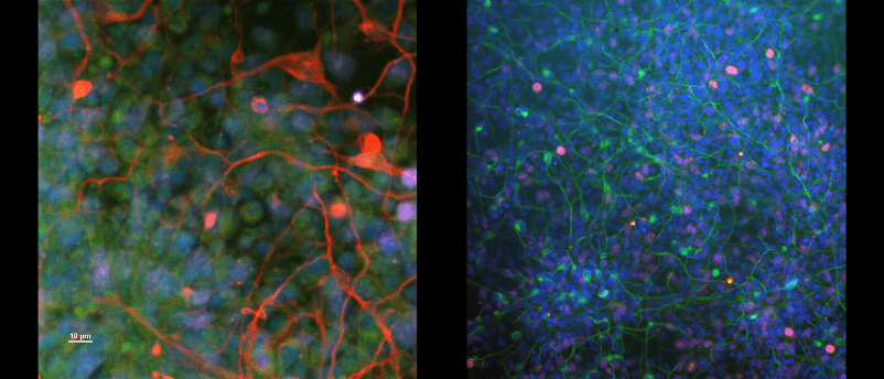
Both images show developing neurons from children with a neurodevelopmental disorder called Smith Lemli Opitz Syndrome.
Credits:
Left: Qutub Lab, Imaging: Dr. Amina Ann Qutub, Cell culture: Dr. Arun Mahadevan. Stem cells are a gift from Dr. Kevin Francis, The Children’s Health Research Center at Sanford Research Institute. Blue = nuclei. Green = Nestin, a neural stem cell marker. Red = MAP2, microtubule-associated protein 2, a cytoskeletal marker for neurons. Purple = Ki67, a proliferative marker. Contrast enhanced. Nikon A1 confocal microscope, 20X.
Right: Qutub Lab, Imaging/cell culture: Dr. Arun Mahadevan. Stem cells are a gift from Dr. Kevin Francis, The Children’s Health Research Center at Sanford Research Institute. Blue = nuclei. Green = TUJ1, a cytoskeletal marker for neurons. Pink = Ki67, a proliferative marker. Contrast enhanced. Nikon Ti-E2 fluorescence microscope, 10X.
What are the best aspects of your job?
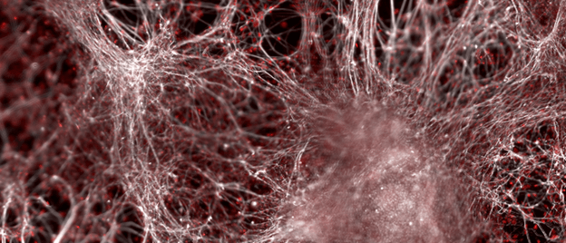
Knowing that my lab’s work will improve lives is an incredible motivator. Another is the sheer joy of scientific discovery and observation. Looking at long-time-lapse imaging of human cells and tissue is mesmerizing. Brain cells are incredibly beautiful, strange and dynamic. Being able to describe how they evolve computationally is like solving a very complex three-dimensional puzzle – you cheer when it is completed.
Another rewarding aspect of my job is working with skilled, passionate students and collaborators from around the world, who enrich our work through diverse perspectives.
What are the most challenging parts?
The most challenging part is knowing that my work is not fast enough. The work in the field (academia, industry and government) is neither fast nor deep enough. It was not fast enough to help my father fully recover from his stroke. Not fast enough for the families who reach out to me for help. My young relative has a pediatric neurodevelopmental disorder, and he said we will eventually understand the brain well enough to know what causes the disorder, but he wasn’t sure this knowledge would come in his lifetime. I assured him we would know in his lifetime, and that I would do whatever it takes.
The time for discovery and a therapeutic breakthrough weighs on me.
What advice would you give to your younger self?
My advice would be:
- Brace yourself. At 20, I had no clue what I would soon face. In my second year in graduate school, I lost my younger sister. Her loss devastated my family and me. Pouring myself into science helped me focus. I sometimes pretended my sister could see what I could. I sought out beauty and patterns in everything I read and saw. Ever since, every difficult period I’ve been through, solving problems in biomedicine – observing, imaging, deep reading, programming or writing – has helped me get through it.
- Surround yourself with people who are open-minded. This seems like obvious advice, but there is no sniff test for open-mindedness. In science, ‘curiosity’ is the trait to look for in fellow researchers, entrepreneurs and inventors. People who are constantly seeking to learn, grow and try new things, who are curious and ask open-ended questions. Those are the people that have most helped my work advance and I most enjoy working with.
- “Whatever you’re meant to do, do it now. The conditions are always impossible.” – Doris Lessing, Nobel Laureate
What resources/networks have you found useful in your career, particularly as a woman in science?
Networks have been transformative for my career, and most emerged out of my curiosity. In graduate school, when I was preparing for my doctoral qualifying exams, I reached out and called all the lead authors of the articles I really liked and asked them a series of questions about the study. I did this out of scientific curiosity, and likely to feel more connected after the loss of my sister.
In retrospect, I am grateful anyone answered – let alone all those who did! It was a wonderful way to learn science. Not only did I learn new techniques, but I also quickly met luminaries in my field through stories, gained a history of the discovery of the blood–brain barrier and secured invitations to share my work.
The habit of reaching out to people whose work inspires me has stayed with me, strengthened with time and spread across domains and countries. Conferences have made the cold calls less necessary and awkward. Most of my collaborations – even those with researchers across the street – have started because of meeting at a conference.
These spontaneous networks augment yet do not replace mentors and friends who have advocated for me. On a daily basis, progress towards broader scientific and biomedical goals is not always linear or obvious. Especially for projects that cross many domains and break traditional ways of doing science, where there are inherent risks and limited resources.
When you are an outlier in your career in other ways too (e.g., of a different gender or race), the best advice I have is to build a network revolving around what you value.