Surveying the mitochondrial machinery
A new comprehensive classification map of mitochondrial proteins promises new insights into mitochondrial function.
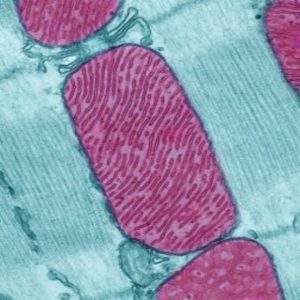
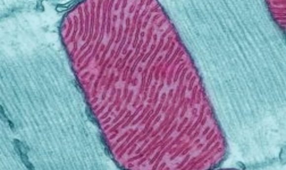
Credit: NIH image gallery
Mitochondria are known as the cell’s “powerhouses” or “generators” for good reason: these small, specialized organelles are responsible for making adenosine triphosphate (ATP), the biological currency of cellular energy. Mitochondrial failure can lead to a host of medical issues affecting the heart, brain, and other organ systems with high energy needs. To date, finding the root causes of (and potential treatments for) mitochondrial disease has been hampered by our lack of understanding of the organelles themselves—including the specific proteins that help them carry out their biological jobs.
“Mitochondria have four subcompartments and each one is made up of specific proteins. In fact, there are more than 800 mitochondrial proteins in yeast and more than 1500 in humans. But it has been a challenge to determine which proteins are responsible for which subcompartments,” said Chris Meisinger, a researcher at the University of Freiburg. “Many of the standard protein databases we have on sublocalization of mitochondrial proteins are incomplete or incorrect.”
Those four subcompartments include the inner and outer portions of the specialized mitochondrial membrane, the intermembrane space, and the matrix, or interior chamber of the organelle. Previous attempts to map the landscape of subcompartmental proteins and their functions left missing or misattributed protein building blocks, so Meisinger and colleagues turned to a unique integrated approach to examine, classify, and map the submitochondrial proteins in S. cerevisiae.
Using a painstaking combination of techniques, the researchers isolated mitochondria from yeast by centrifugation, collected proteins from each of the four compartments using sonication, extracted the membrane proteins using carbonite methods, and distinguished between the inner and outer membraned by comparing the extracted vesicles. With the help of a mass spectrometry, isotope labeling, and a homegrown statistical model, Meisinger’s team then mapped the full protein landscape for mitochondria. In doing so, they created the first complete quantitative map of protein distribution for yeast mitochondria—so complete that the group successfully reassigned proteins whose location had been in question prior to this work.
“Mitochondria have many important functions and are critical to cell health. Understanding which protein belongs to which subcompartment is very important so we can better understand the metabolic pathways as well as help us discover the function of new proteins,” said Meisinger. “This may one day help us better understand what goes wrong in mitochondrial disease as well as help develop new interventions to treat it.”