Docs unlock the pox: key insights into the molecular structure of poxvirus cores
Researchers utilize electron microscopy coupled with AlphaFold software to glean key insights into the structure of the core of poxviruses.
Despite the global effort to eradicate Smallpox, achieved in 1980, poxviruses remain a serious threat to public health. Over the past 2 years, a resurgence of monkeypox (or Mpox) has been reported worldwide, an ever-present cause for concern.
In order to treat viral infections, and prevent future outbreaks, a thorough understanding of the virus is essential; this includes the vectors of its transmission and its molecular structure, which is particularly vital when developing drugs or vaccines to combat them.
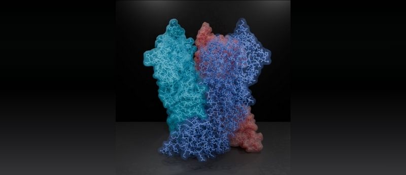
The protein A10, common to all clinically relevant poxviruses, has represented a particular blind spot in our understanding of the function and pathology of these viral strains. A10 is a central piece in the core of the poxvirus. A new study from researchers at the Institute of Science and Technology Austria (ISTA) (Klosterneuburg, Austria), has finally shed light on this linchpin protein, expanding the field and potentially leading the way for future therapeutics against poxviruses.
Florian Schur, corresponding author of the paper, explained the central aim of the study, “we know that for poxviruses to be infective, their viral core must be properly formed. But what is this poxviral core made of, and how do its individual components come together and function?” Together with his co-authors, Schur utilized a range of cutting-edge experimental and computational techniques to probe the structure and function of the poxvirus core.
Despite growing interest in the structure of poxvirus core proteins, there remains a drought of experimentally determined structures. The team utilized the revolutionary AlphaFold software, which has the ability to predict the tertiary structure of proteins from their primary amino acid sequence. Once this was achieved, it was time for the virus to go under the microscope.
Mature Vaccinia virions, primary components of the smallpox vaccine, and isolated poxviral cores were subjected to an array of microscopy techniques. “We combined the ‘classic’ single-particle cryogenic electron microscopy (cryo-EM), cryo-electron tomography, subtomogram averaging, and AlphaFold analysis to gain an overall view of the poxviral core”, explains co-author Julia Datler. Cryo-EM is a tried and tested method of ascertaining the structure of complex biomacromolecules in their active states. It has advantages over crystallography by allowing the protein to move freely until flash-frozen into place, whereas crystallography typically opts for the most stable conformation. However, this can make reconstructing the 3D structure of the protein a challenge.
Cryo-EM reveals new structural elements in a key ciliary protein complex
Scientists capture the protein complex responsible for transporting signaling molecules to cilia in unprecedented detail.
Cryo-electron tomography, a ‘specialty’ of the Schur group, is a technique not dissimilar to the routine CT scans employed in clinical settings, only on a much smaller scale. The technique is carried out by imaging the protein at different angles, titling the sample during the experiment. The resulting images can then be compiled to create 3D representations of the protein, in nanometer resolutions.
The results allowed the group to fit the AlphaFold model into experimentally observed shapes and identify critical molecules in the structure of the poxvirus core. “We found that A10 defines key structural elements of the core of poxviruses”, Datler commented.
It cannot be understated how difficult an undertaking probing the structure of a complex integration of biological macromolecules can be, as on the nanoscale the extraordinary complexity of the natural world is laid bare. Proteins are gigantic, unwieldy molecules in constant motion, yet for us to understand their structure or their function, myriad factors must align to create a snapshot of the system in fleetingly perfect order.
The team is confident that their findings will enrich the field of poxvirus study. “These findings are a great resource to interpret bits of structural and virological data generated over the last decades” says Schur. Looking forward, Schur adds that the results could be used to design therapeutics targeted to the core of the poxvirus, “one could think of drugs that prevent the core from assembling – or even disassembling and releasing their viral DNA during infection. Ultimately, fundamental virus research, as done here, allows us to be better prepared against possible future viral outbreaks.”