Da Vinci heart mystery settled with imaging genetics
A 16th Century mystery about the trabeculae of the heart has been solved with the power of data, AI and imaging genetics.
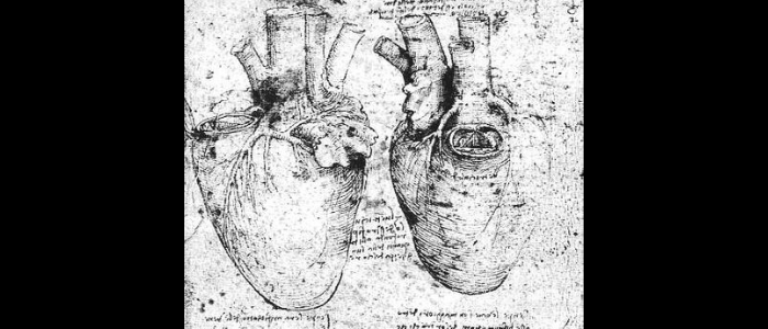
500 years ago, Leonardo da Vinci sketched heart trabeculae – the intricate network of muscle fibers that form geometric patterns on the inner surface of the heart – which he speculated warmed the blood. While now known to help oxygenate the heart during development, their function in adults had remained a mystery – until new results from a multidisciplinary team comprising researchers from institutions including EMBL’s European Bioinformatics Institute (EMBL-EBI; Cambridge, UK), Cold Spring Harbor Laboratory (NY, USA) and the MRC London Institute of Medical Sciences (UK).
The research utilized MRI, artificial intelligence (AI) and genetic data from 25,000 people in the UK biobank and 50,000 patients with heart failure. Speaking to BioTechniques, corresponding author Declan O’Regan (MRC London Institute of Medical Sciences), explained: “Trabeculae line the inner surface of the heart and form a complex network of muscles – so we suspected that they had an important role in how the heart pumps. We analyzed each cardiac MRI using fast AI algorithms and then searched for genetic variants linked to geometric patterns in these structures. We also looked at 50,000 patients with heart failure to show that they affect your chances of having disease.”
Data from the UK Biobank was key to the research, with collaborators at Imperial College London (UK), led by O’Regan, deriving a new machine learning algorithm to automatically measure the degree of trabeculation from the cardiac MRIs.
 Cordio HearO: the app that could detect heart failure by the sound of your voice
Researchers have developed a speech analysis smartphone app, called Cordio HearO, with the ability to detect acute heart failure.
“The power of UK Biobank is its wealth of genetic and phenotype data on a large number – 500,000! – of participants,” Principle Investigator Hannah Meyer (Cold Spring Harbor Laboratory) explained to BioTechniques. “At the same time, the large amount of data poses challenges in their analyses. Only through sophisticated machine learning methods and the use of powerful computers on which we could conduct 1000s of analyses in parallel could we conduct our research.” The team has made their analysis code freely available on GitHub.
Meyer’s group provided the genetics expertise, looking for genetic variants linked to the structures to determine their effect on heart disease risk.
The results suggest the rough surface allows blood to flow more efficiently, with different fiber patterns affecting the risk of heart failure. Six DNA regions were highlighted that affect the patterns, two of which are also involved in the branching of nerve cells, implying a similar mechanism could occur in the developing brain.
“Heart failure affects up to 2% of the population and has a poor outcome,” O’Regan noted. “This work reveals a previously unknown mechanism determining heart function and future research will explore whether this can identify new approaches for prevention and treatment.”
In addition to linking trabeculae to heart disease, the study exemplifies the power of multidisciplinary research and imaging genetics. “Imaging genetics uses computational algorithms to extract features from medical images to subsequently study the link between these features and genetics. With the wealth of medical imaging data in UK Biobank, there are many other exciting opportunities to use imaging genetics to study organ function,” noted Meyer.