Neural networks in a dish
In the latest development for the rapidly evolving field of cerebral organoids, a team of researchers has created a fully functional neural network, derived from 3D cell cultures.
Whether you call them ‘mini-brains’ or ‘brains-in-a-dish’, cerebral organoids are taking over the research world and are a novel way to model human development and disease. In a recent development for the field, researchers from Japan have created in vitro fully functional neural networks that are derived from cerebral organoids.
The potential for organoids is vast with applications across the fields of medical research and drug discovery, “because they can mimic cerebral development, cerebral organoids can be used as a substitute for the human brain to study complex developmental and neurological disorders,” explained corresponding author Jun Takahashi of Kyoto University (Japan).
However, as research stands currently, organoids are limited tools as they lack all of the necessary supporting structures that are present in their in vivo counterpart. Without the complex vasculature and blood–brain barrier you are restricted in what conclusions you are able to draw from the models, making it difficult to comprehensively evaluate their neuronal network function. This is a difficultly the Japanese team hope to have overcome in their latest networks.
“In our study, we created a new functional analysis tool to assess the comprehensive dynamic change of network activity in a detected field, which reflected the activities of over 1,000 cells,” commented first author Hideya Sakaguchi, previously a fellow at Kyoto University, now working at the Salk Institute (CA, USA). “The exciting thing about this study is that we were able to detect dynamic changes in the calcium ion activity and visualize comprehensive cell activities.”
-
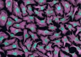
Mini-brains created from primary cells
-
Talking Tech News | Cerebral organoids: past, present, future
-
Grow your own brain
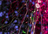
To create their networks, Sakaguchi et al. generated a ball of pluripotent stem cells that they then placed into a dish filled with a culture medium optimized to mimic the environment needed for cerebral development. The team could visualize synchronized and non-synchronized activity within the network as well as individual connections between distinct neurons.
Sakaguchi hopes that this model will help fellow researchers to understand the neural processes by which information is encoded in the brain, as well as the underlying mechanisms of psychiatric disease. “We believe that our work introduces the possibility of a broad assessment of human cell-derived neural activity,” he added.
As research into cerebral organoids grows, so does the ethical concern as other researchers raise the question of consciousness. “Because cerebral organoids mimic the developmental process, a concern is that they also have mental activities such as consciousness in the future,” Sakaguchi explained.
“Some people have referenced the famous ‘brains in a vat’ thought experiment proposed by Hilary Putnam, that brains placed in a vat of life-sustaining liquid with connection to a computer may have the same consciousness as human beings.”
That being said, the team doubt that consciousness will ever develop in these cellular models as they lack input from the environment and their surroundings; “consciousness requires subjective experience, and cerebral organoids without sensory tissues will not have sensory input and motor output,” argued Sakaguchi.
“However, if cerebral organoids with an input and output system develop consciousness requiring moral consideration, the basic and applied research of these cerebral organoids will become a tremendous ethical challenge,” he added.
Takahashi believes that future research into cerebral organoids will be focused in three key areas: drug discovery, regenerative medicine and modeling neuropsychiatric disorders. “Cerebral organoids can bring great advances to pharmacological companies by replacing traditional animal models and can also be used to model untreatable neural diseases,” he added.
“Using our method, it will be possible to analyze cell activity patterns in brain functions to further explore these areas.”