Cryo-electron microscopy takes the chemistry Nobel
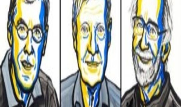
The 2017 Nobel Prize in Chemistry is awarded to three researchers for the development of cryo-electron microscopy.
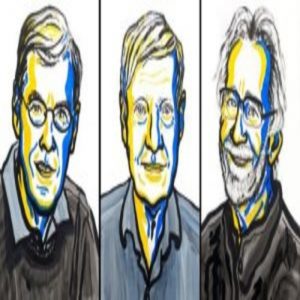
Credit: Niklas Elmehed, Nobel Media 2017
This morning, the Royal Swedish Academy of Sciences announced that the 2017 Nobel Prize in Chemistry was awarded jointly to Jacques Dubochet of the University of Lausanne in Switzerland, Joachim Frank of Columbia University in New York, and Richard Henderson from the MRC Laboratory of Molecular Biology in Cambridge for their groundbreaking work in developing the technique of Cryo-electron microscopy (cryo-EM).
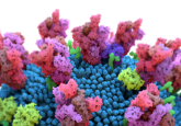
Cryo-EM is an important method for determining the structures of biological molecules at high-resolution. Unlike traditional electron microscopy, cryo-EM does not require sample fixation. And by using extremely low temperatures to freeze biomolecules, the technique even allows researchers to visualize biological processes.
Initially, a major challenge associated with the concept of cryo-EM was that crystalline ice formed during the cooling process, leading to diffraction and imaging problems during electron microscopy. In 1981, Jacques Dubochet demonstrated a way to cool water rapidly for use in cryo-EM. Dubochet and his team placed water onto a grid that had been immersed in liquid propane at -190oC. That layer of water yielded uniform absorption of electrons in the cryo-EM. Dubochet noted that if kept below -160oC, it would be possible to solidify the water around a biomolecule within an electron microscope, creating the opportunity to image the shape of a biological sample within a solution. Dubochet went on to do just this in 1984 when his group presented cryo-EM images of viruses in suspension.
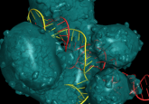
On the heels of Dubochet’s work, Richard Henderson took cryo-EM one step further in 1990. Henderson used the technique to generate high-resolution images of individual biomolecules, such as proteins, using cryo-EM. His idea was to average thousands of images to reconstruct a protein structure. Henderson and his team applied their cryo-EM approach to bacteriorhodopsin, showing that it was possible to limit radiation damage for a biological sample while still retaining information on the positions of side-chains within the protein. It was this breakthrough that, according to the Nobel committee, “proved the technology’s potential.”
Joachim Frank was cited in the Nobel announcement for his efforts between 1975 and 1986 to make cryo-EM more generally accessible to the scientific community. His team developed image processing methods that yielded three-dimensional structures of biomolecules by merging the fuzzy two-dimensional images obtained in cryo-EM when using non-destructive electron doses. This work led to a suite of computer tools that researchers can now use for the analysis of structures based on cryo-EM datasets.
The laureates will share the 9 million Swedish krona prize.