Three-dimensional residual channel attention networks denoise and sharpen fluorescence microscopy image volumes
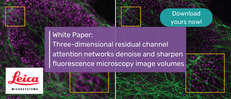
Aivia scientists and their collaborators at the NIH and the University of Maryland (MD, USA) have developed a 3D residual channel attention network (3D RCAN) that denoises and/or improves the spatial resolution of fluorescence-microscopy image volumes with a performance that is competitive to state-of-the-art neural networks.
This achievement is a critical contribution for improving both fluorescence-microscopy data and gold-standard alternatives that highlights the power of using a single platform like Aivia software to unlock insights from complex image datasets.
Download your White Paper now.
Download White PaperMore information
In this White Paper, discover:
- How to utilize the 3D RCAN models
- The challenges of fluorescent microscopy
And much more!
This content was provided by Leica.